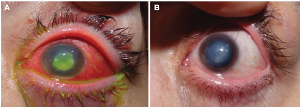
What is a corneal ulcer ?
A corneal ulcer is an open sore or epithelial defect with underlying inflammation on the cornea, the clear structure in the front of the eye. The cornea overlies the iris, which is the colored part of the eye and is separated from the iris by the aqueous fluid in the anterior chamber of the eye.
Most corneal ulcers are caused by infections and can be bacterial (common in people who wear contact lenses), viral herpes simplex virus and varicella virus, or fungal (improper care of contact lenses or overuse of eyedrops that contain steroids). Symptoms include red eyes, pain, feeling like something is in the eye, tearing, pus/thick discharge, blurry vision, pain from bright lights, swollen eyelids, or a white or gray round spot on the cornea.
How does a dr.sandeep arora diagnose a corneal ulcer?
The presence of a corneal ulcer can be diagnosed by an ophthalmologist (and other medical caregivers) through an eye examination. The ophthalmologist will be able to detect an ulcer by using a special eye microscope known as a slit lamp. A drop containing the dye fluorescein, when placed in the eye, can make the ulcer easier to see. Scrapings of the ulcer may be sent to the laboratory for identification of bacteria, fungi, or viruses. Certain bacteria, such as a species of Pseudomonas, may cause a corneal ulcer which is rapidly progressive.
What are corneal ulcer treatment options?
Treatment is aimed at eradicating the cause of the ulcer. Anti-infective agents directed at the inciting microbial agent will be used in cases of corneal ulcer due to infection. Generally, these will be in the form of drops or ointments to be placed in the eye; but occasionally, especially in certain viral infections, oral medications will also be employed. Occasionally, steroids will be added, but should only be used after examination by an eye doctor or other physician using a slit lamp, because in some situations, steroids may hinder healing or aggravate the infection. Subconjunctival injection of antibiotics may occasionally be utilized.
In cases of patients aggravated by eye dryness or corneal exposure (for example, corneal exposure to a dry and/or sandy environment), tear substitutes will be used, possibly accompanied by patching or a bandage contact lens.
In corneal ulcers involving injury, the inciting agent must be removed from the eye (using copious irrigation for chemicals or by using a slit lamp microscope to remove particles such as wood or metal) and then adding medications to prevent infection and minimize scarring of the cornea.
If the corneal ulcer is due to an eyelash growing inward, the offending lash should be removed, together with its root. If it grows back in an abnormal manner, the root may have to be destroyed using a low-voltage electrical current. If the corneal ulcer is secondary to the eyelid turning inward, surgery directed at correctly repositioning the eyelid may be necessary.
Contact lenses should be discontinued in the affected eye of any case of corneal ulcer, regardless of whether the ulcer was initially caused by the contact lens.
If the ulcer cannot be controlled with medications, it may be necessary to surgically debride the ulcer. If the ulcer causes significant corneal thinning and threatens to perforate the cornea, a surgical procedure known as a corneal transplant (keratoplasty) may be necessary.
Individuals with corneal ulcers due to immunological diseases may require patient-specific treatment with immunosuppressive drugs. Such patients may require care coordinated with an ophthalmologist in conjunction with other doctors. In patients with secondary iritis associated with a corneal ulcer, cycloplegic eyedrops may be used to decrease pain and dilate the pupil.
Anyone with an irritated eye that does not improve quickly after removing a contact lens or after mild irrigation should contact an ophthalmologist immediately. Do not borrow someone's eyedrops.

 +91 982-577-8009